Senin, 18 April 2011

Fisika Kedokteran

VISION

  1. To be a center of excellence in physics and applied physics research and education in the national and international level
  2. To produce world-class graduates who are highly innovative and competent in scientific, academic and industrial environments.

Pengantar

Fisika berasal dari bahasa Yunani, physikos yang berarti ‘alamiah’. Karena itu Fisika merupakan suatu ilmu pengetahuan dasar yang mempelajari gejala-gejala alam dan interaksinya yang terjadi di alam semesta ini. Hal-hal yang dibicarakan di dalam fisika, selalu didasarkan pada pengamatan eksperimental dan pengukuran yang bersifat kuantitatif.

Fisika terbagi atas dua bagian yaitu :

1. Fisika klasik yang meliputi bidang : Mekanika, Listrik Magnet, Panas,Bunyi, Optika dan Gelombang.

2. Fisika Modern adalah perkembangan Fisika mulai abad 20 yaitu penemuan Relativitas Einsten. (Atom Inti, Radiasi dll)

Ilmu Fisika mendukung perkembangan teknologi, enginering (teknik sipil, elektro, mesin, pertambangan, dll), bahkan di bidang kesehatan (mis: kebidanan, keperawatan, kedokteran) dan lain-lain.

Dalam kesehatan kajian Fisika, seringkali dicirikan dengan awalan Bio: Biomekanika, Biothermal, Biofluida, Bioakustik, Biolistrik, Biooptik, Bioradiasi, dan lain-lain. :)

[ Untuk materi dasar-dasar Fisika tentang Besaran Pokok - Turunan, Satuan, Vektor dan Skalar dapat dilihat di Materi Ringkas Fisika Dasar. ]
tubuh01


Berikut ini adalah materi tambahan terkait aspek Kesehatan.

Pengukuran Parameter Fisik

Berikut ini disajikan data standar manusia yang menggunakan sistem satuan internasional, turunan SI dan non SI untuk umur 30 tahun.

tab00

Dapat dicermati bahwa satuan dari besaran-besaran yang lazim digunakan dalam pengukuran fisika cukup familiar di gunakan dalam mempelajari fisika kesehatan.

Berikut ini contoh terapan lainnya:

Indeks Massa Tubuh:

yaitu pengukuran antropometri untuk menilai postur tubuh ideal atau apakah komponen tubuh sesuai dengan standard normal atau ideal. Pengukuran antropometri yang paling sering digunakan adalah rasio antara berat badan (dalam kg) dan tinggi badan (dalam m) kuadrat, yang disebut Indeks Massa Tubuh (IMT) sebagai berikut :

IMT = BB (kg) / (TB)^2
—————————————–catatan :) ————
Anda akan temui kesalahfahaman penggunaan besaran berat, dalam Berat Badan (BB) yang seharusnya adalah massa badan, dengan penggunaan satuan kg ( yang ini sudah benar). Di masyarakat sendiri, hal tersebut seringkali terjadi…. perhatikan di pasar2, :)
—————————————————————-
tab01

obesitas perlu diwaspadai karena biasanya berpotensi juga menderita penyakit degeneratif seperti Diabetes Melitus, hipertensi, hiperkolesterol dan kelainan metabolisme lain yang memerlukan pemeriksaan lanjut baik klinis atau laboratorium

Disebutkan bahwa batas ambang normal untuk laki-laki adalah: 20,1–25,0; dan untuk perempuan adalah : 18,7-23,8. Untuk kepentingan pemantauan dan tingkat defesiensi kalori ataupun tingkat kegemukan, lebih lanjut FAO/WHO menyarankan menggunakan satu batas ambang antara laki-laki dan perempuan. Ketentuan yang digunakan adalah menggunakan ambang batas laki-laki untuk kategori kurus tingkat berat dan menggunakan ambang batas pada perempuan untuk kategorigemuk tingkat berat.

Untuk kepentingan Indonesia, batas ambang dimodifikasi lagi berdasarkan pengalam klinis dan hasil penelitian dibeberapa negara berkembang. Pada akhirnya diambil kesimpulan, batas ambang IMT untuk Indonesia adalah sebagai berikut:
tab02

Jika seseorang termasuk kategori :
1. IMT 27,0 : keadaan orang tersebut disebut gemuk dengan kelebihan berat badan tingkat berat

Contoh cara menghitung :

Opong dengan tinggi badan 159 cm, mempunyai berat badan 70 kg. Maka IMT Opong adalah :
70 70
——————– = ——– = 27,7
(1,59 X 1,59) m 2,53
Berarti status gizi Opong adalah gemuk tingkat berat, dan Opong dianjurkan menurunkan berat badannya sampai menjadi 47- 63 kg agar mencapai berat badan normal (dengan IMT 18,5 – 25,0).

PERHATIAN !
Seseorang dengan IMT > 25,0 harus berhati-hati agar berat badan tidak naik. Dianjurkan untuk menurnkan berat badannya sampai dalam batas normal.

Berat Badan Ideal :
Atau untuk mengetahui Berat Badan ideal dapat menggunakan rumus Brocca sebagai berikut :
BB ideal = (TB – 100) – 10% (TB – 100)
Batas ambang yang diperbolehkan adalah + 10%. Bila > 10% sudah kegemukan dan bila diatas 20% sudah terjadi obesitas.
Contoh: wanita dengan TB = 161 cm, BB = 58 kg
BB ideal = (161 – 100) – 10% (161 – 100)
= 61 – 6,1 = 54,9 (55 kg)
BB 58 kg masih dalam batas > 10%.

Lila (Lingkar Lengan Kiri Atas):
Pengukuran lain yang dapat dilakukan untuk menilai apakah seseorang tersebut kurus menderita kurang gizi, normal atau gemuk, dengan mengukur Lingkar lengan kiri atas (Lila). Biasanya dilakukan pada wanita usia 15 – 45 tahun. Bila Lila < 23,5 cm, wanita tersebut menderita Kurang Energi Kronis (KEK).

RLPP (Rasio Lingkar Perut dan Lingkar Pinggang):
Pengukuran antropometri lain yang sering digunakan adalah mengukur rasio Lingkar perut dan Lingkar Pinggang (RLPP). Pada wanita RLPP yang disarankan < 0,8 sedangkan pada laki-laki 0,8 pada wanita dan > 1 pada laki-laki mempunyai risiko menderita penyakit jantung lebih besar dari yang RLPP nya dibawah ambang batas.

3 Hal yang menjadi faktor penentu ketepatan tindakan pengukuran, yaitu: (1) Alat, (2) Metode, dan (3) manusia-nya.

Kesalahan Pengukuran dalam Tindakan Medis (False)
Untuk menyatakan seseorang sakit atau tidak, perlu dilakukan pengukuran terhadap besaran fisis tubuh seperti suhu badan, tekanan darah, frekuensi detak jantung dan sebagainya.

Kesalahan dapat terjadi dari proses pengukuran disebabkan: faktor alat, metode maupun human error, sehingga terjadi false positif dan negatif.

False Positif: Error yang terjadi dimana penderita dinyatakan menderita suatu penyakit padahal tidak
False Negatif: Error yang terjadi dimana penderita dinyatakan tidak sakit padahal menderita suatu penyakit

Untuk menghindari :
1. Dalam pengambilan pengukuran
2. Pengulangan pengukuran
3. Penggunaan alat yang dapat dipercaya
4. Kalibrasi terhadap alat.

Sumber:

http://alifis.wordpress.com/2009/10/24/seri-fisika-kesehatan_pendahuluan/

(Alifis@Corner)

Jumat, 18 Maret 2011

Nuclear medicine

Nuclear medicine is a branch or specialty of medicine and medical imagingthat uses radionuclides and relies on the process of radioactive decay in the diagnosis and treatment of disease.


In nuclear medicine procedures, elemental radionuclides are combined with other elements to form chemical compounds, or else combined with existingpharmaceutical compounds, to form radiopharmaceuticals. These radiopharmaceuticals, once administered to the patient, can localize to specific organs or cellular receptors. This property of radiopharmaceuticals allows nuclear medicine the ability to image the extent of a disease-process in the body, based on the cellular function and physiology, rather than relying on physical changes in the tissue anatomy. In some diseases nuclear medicine studies can identify medical problems at an earlier stage than other diagnostic tests.

Treatment of diseased tissue, based on metabolism or uptake or binding of a particular ligand, may also be accomplished, similar to other areas of pharmacology. However, the treatment effects of radiopharmaceuticals rely on the tissue-destructive power of short-range ionizing radiation.


Further reading

  • Mas JC: A Patient's Guide to Nuclear Medicine Procedures: English-Spanish. Society of Nuclear Medicine, 2008. ISBN 978-0972647892
  • Taylor A, Schuster DM, Naomi Alazraki N: A Clinicians' Guide to Nuclear Medicine, 2nd edition. Society of Nuclear Medicine, 2000. ISBN 978-0932004727
  • Mark J. Shumate MJ, Kooby DA, Alazraki NP: A Clinician's Guide to Nuclear Oncology: Practical Molecular Imaging and Radionuclide Therapies. Society of Nuclear Medicine, January 2007. ISBN 978-0972647885
  • Ell P, Gambhir S: Nuclear Medicine in Clinical Diagnosis and Treatment. Churchill Livingstone, 2004. (1950 pages) ISBN 978-0443073120
External links

Jumat, 18 Februari 2011

Photomedicine

Photomedicine is an interdisciplinary branch of medicine that involves the study and application of light with respect to health and disease. Photomedicine may be related to the practice of various fields of medicine including dermatology, surgery, interventional radiology, optical diagnostics, cardiology, and oncology.

A branch of photomedicine is light therapy.


Examples

See also

External links

Journals and Societies

Miscellaneous

Selasa, 18 Januari 2011

Interventional radiology

Interventional radiology (abbreviated IR or sometimes VIR for vascular and interventional radiology, also known as Image-Guided Surgery or Surgical Radiology) is a subspecialty of radiology, or in Europe a separate subspecialty of medicine in its own right, in which minimally invasive procedures are performed using image guidance. Some of these procedures are done for purely diagnostic purposes (e.g., angiogram), while others are done for treatment purposes (e.g., angioplasty).

The basic concept behind interventional radiology is to diagnose or treat pathology with the most minimally invasive technique possible. Images are used to direct interventional procedures, which are usually done with needles and narrow tubes called catheters. The images provide road maps that allow the interventional radiologist to guide these instruments through the body to the areas containing disease. By minimizing the physical trauma to the patient, peripheral interventions can reduce infection rates and recovery time as well as shorten hospital stays.[1]

History

The advancements in the field of radiological imaging such as the Seldinger technique, together with innovations in instrumentation, led to a rapid development in interventional procedures in the 1970s.Cardiovascular procedures were found to be particularly well-suited for guided and minimally invasive operations, and catheterization remains as one of the main applications for interventional radiology.

Nobel nominee Charles Dotter is considered the "father of angioplasty and interventional radiology".[2]

See also: Interventional cardiology


Training

As in most medical specialties, training varies depending on varying rules and regulations from country to country. In the United States, interventional radiologists are physicians whose education and training traditionally includes completing a college degree, four years of medical school, a year of training in general medicine and/or surgery (internship), a four year diagnostic radiology residency program, and then a one or two year fellowship in vascular & interventional radiology. Alternative pathways exist.


Imaging Modalities

Common interventional imaging modalities include fluoroscopycomputed tomography (CT), ultrasound (US), and magnetic resonance imaging (MRI). Fluoroscopy and computed tomography use ionizing radiation that may be potentially harmful to the patient and the interventional radiologist. However, both methods have the advantages of being fast and geometrically accurate. Ultrasound suffers from image quality and tissue contrast problems, but is also fast and inexpensive. Magnetic resonance imaging provides superior tissue contrast, at the cost of being expensive and requiring specialized instruments that will not interact with the magnetic fields present in the imaging volume.


Procedures

Common IR procedures are:

  • Angiography: imaging the blood vessels to look for abnormalities with the use of various contrast media, including iodinated contrast, gadolinium based agents, and CO2 gas.
  • Chemoembolization: delivering cancer treatment directly to a tumour through its blood supply, then using clot-inducing substances to block the artery, ensuring that the delivered chemotherapy is not "washed out" by continued blood flow.
  • Cholecystostomy: placement of a tube into the gallbladder to remove infected bile in patients with cholecystitis, an inflammation of the gallbladder, who are too frail or too sick to undergo surgery
  • Drain insertions: placement of tubes into different parts of the body to drain fluids (e.g., abscess drains to remove pus, pleural drains)
  • Embolization: blocking abnormal blood (artery) vessels (e.g., for the purpose of stopping bleeding) or organs (to stop the extra function e.g. embolization of the spleen for hypersplenism) including uterine artery embolization for percutaneous treatment of uterine fibroids. Various embolic agents are used, including alcohol, glue, metallic coils, poly-viny alcohol particles, Embospheres, encapsulated chemo-microsphere, and gelfoam.
  • Thrombolysis: treatment aimed at dissolving blood clots (e.g., pulmonary emboli, leg vein thrombi, thrombosed hemodialysis accesses) with both pharmaceutical (TPA) and mechanical means
  • Biopsy: taking of a tissue sample from the area of interest for pathological examination from a percutaneous or transjugular approach
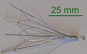
  • IVC filters: - metallic filters placed in the inferior vena cavae to prevent propagation of deep venous thrombus, both temporary and permanent.
Inferior vena cava filter
  • Nephrostomy placement: Placing a catheter directly into the kidney to drain urine in situations where normal flow of urine is obstructed. NUS catheters are nephroureteral stents which are placed through the ureter and into the bladder.
  • Dialysis access and related intervention: Placement of tunneled hemodialysis catheters, peritoneal dialysis catheters, and revision/thrombolysis of poorly functioning surgically placed AV fistulas and grafts.
  • TIPS : Placement of a Transjugular Intrahepatic Porto-systemic Shunt (TIPS) for management of select patients with critical end-stage liver disease and portal hypertension
  • Biliary intervention - Placement of catheters in the biliary system to bypass biliary obstructions and decompress the biliary system. Also placement of permanent indwelling biliary stents.
  • Endovenous laser treatment of varicose veins - Placement of thin laser fiber in varicose veins for non-surgical treatment of venous insufficiency
  • Radioembolization: Embolization of liver with radioactive microspheres of glass or plastic, to kill tumors while minimizing exposure to healthy cells.


Tools

There are a number of catheters used in interventional radiology that can be loosely divided into five types:

  • Diagnostic angiographic catheters
  • Micro catheters
  • Drainage catheters
  • Balloon catheters
  • Central venous catheters


See also


References

  1. ^ Society of Interventional Radiology -- Global Statement Defining Interventional radiology.http://www.sirweb.org/news/newsPDF/IR_Global_Statement.pdf
  2. ^ Lakhan SE, Kaplan A, Laird C, Leiter Y (2009). "The interventionalism of medicine: interventional radiology, cardiology, and neuroradiology"International Archives of Medicine 2 (27): 27. doi:10.1186/1755-7682-2-27PMC 2745361PMID 19740425.
  • Rösch J, Keller F, Kaufman J (2003). "The birth, early years, and future of interventional radiology". J Vasc Interv Radiol 14 (7): 841–53. PMID 12847192.


External links